
Toxicology Triaging System
- Suite of solutions to expedite reporting of preclinical toxicologic pathology studies in rodents.
- Set of algorithms to separate and triage sections from different tissues in toxicology studies.
- Pre-sorted images for triaged reporting.
- Automated organ separation to identify organs and enable batch processing.
- Batch analysis for faster turnaround of high throughput toxicology studies.
- Segmentation and quantification of “Not normal” features.
- Quantitative results with comprehensive display of annotated outputs.
- Compliance with INHAND terminology.
- Accelerated translation and application to additional species or tissue types.
- Efficient workflow resulting in significant reduction in drug discovery time.
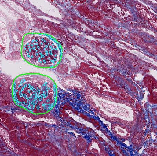
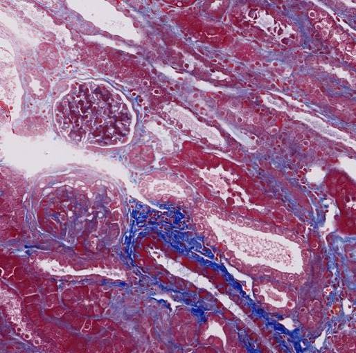
Glomerulosclerosis Quantification
- Quantification of histomorphological parameters in efficacy models for Chronic Kidney Disease .
- Segments glomeruli and quantifies glomerulosclerosis in Masson Trichrome stained sections from rodent kidney .
- Reduces the time and effort expended in manual annotation of glomeruli and quantification of glomerulosclerosis .
- Improves accuracy and reproducibility of quantification results .
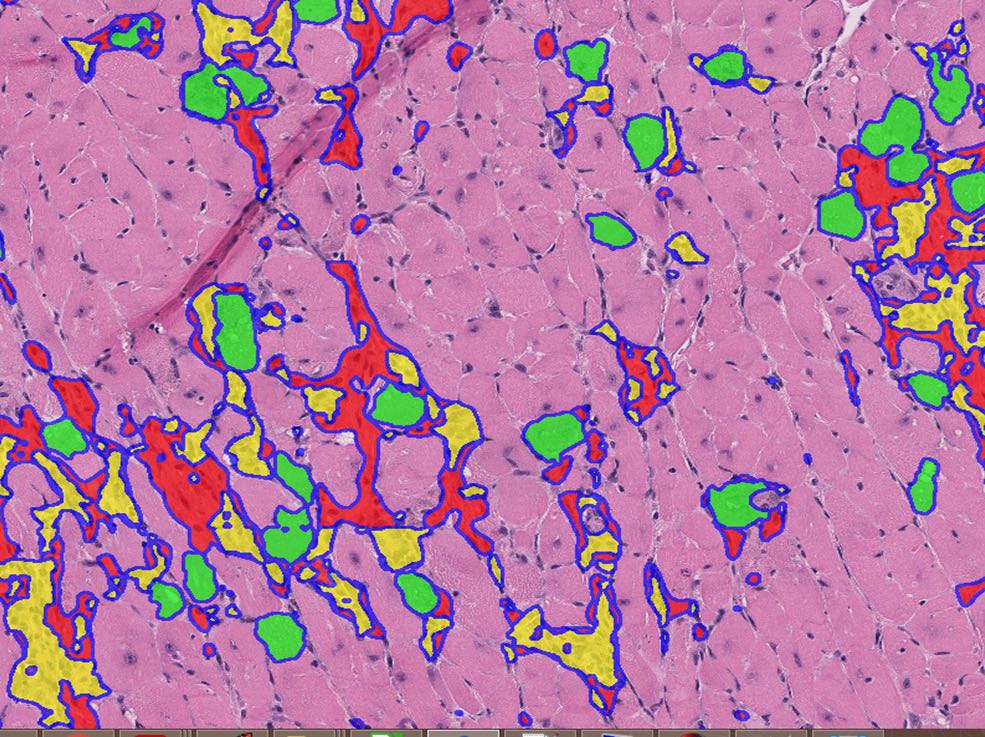
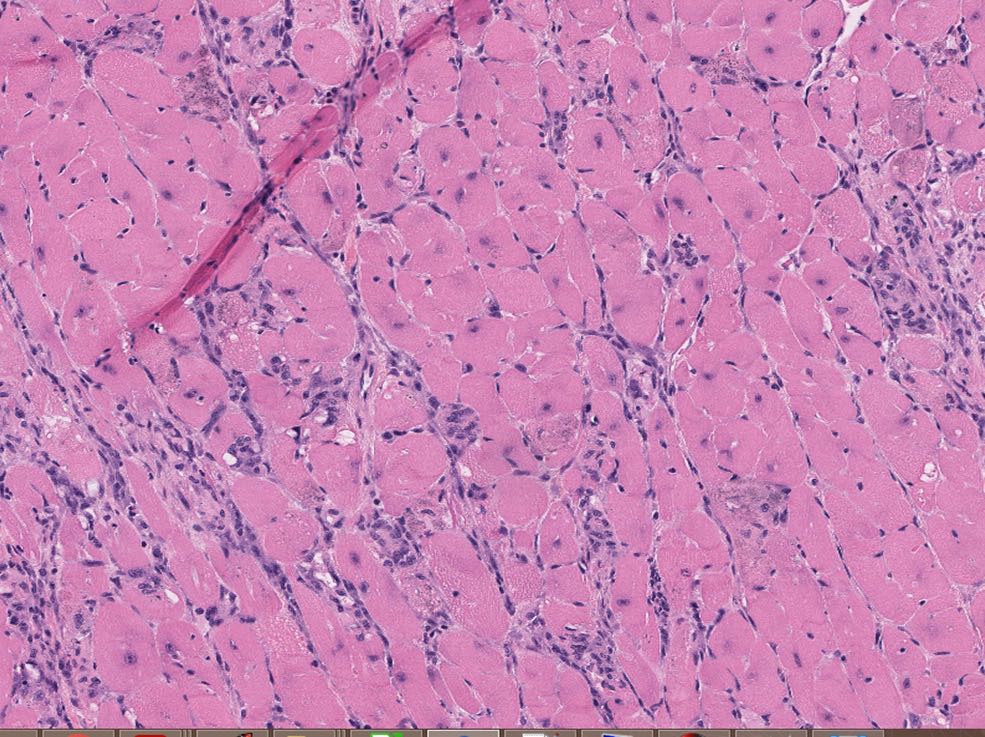
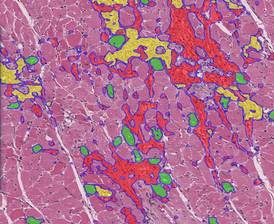
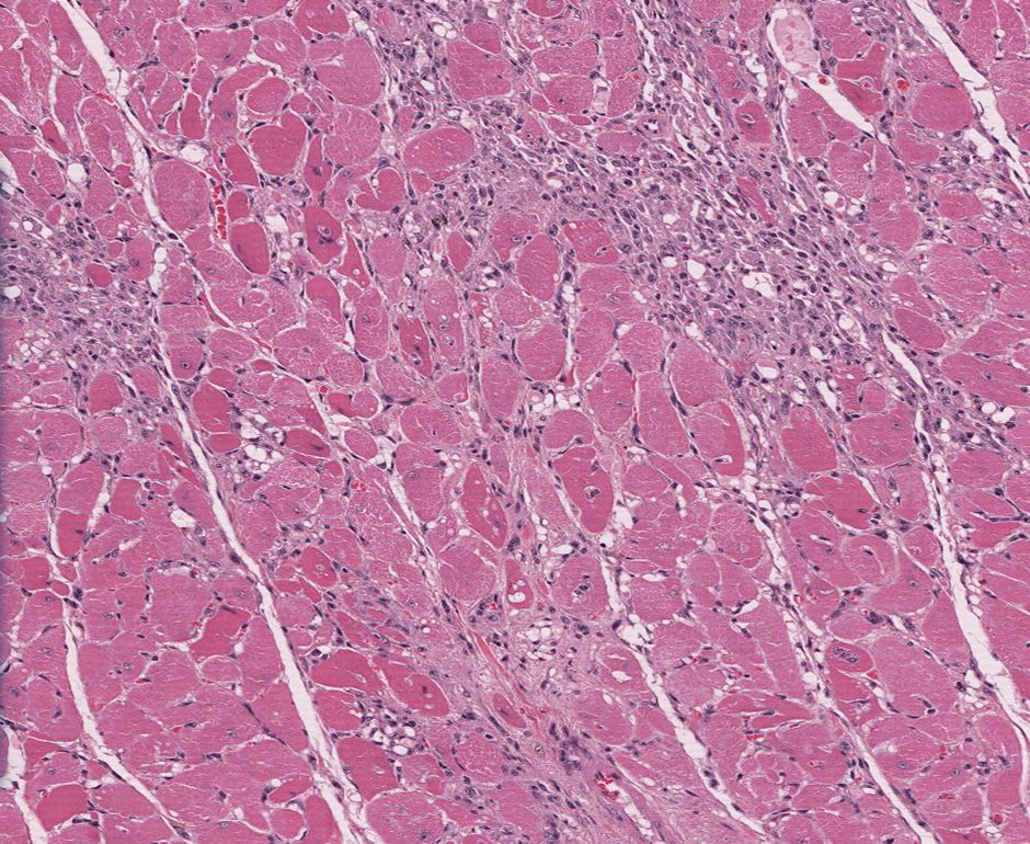
Rodent Cardiomyopathy Scoring
- Solutions for sensitive and consistent detection and quantification of Progressive Cardiomyopathy (PCM) features in rodent heart sections .
- Objective quantification of the histomorphological features of PCM, to assist pathologists in discerning cardiotoxicity-associated changes .
- Accurate severity scoring to help differentiate spontaneous from test article-related findings .
- High values for accuracy, intersection over union, and dice coefficient for each individual feature .
- Strong positive correlation with the median severity grade assigned by panel of pathologists and the percentage area of the heart predicted to have PCM lesions by the algorithm .
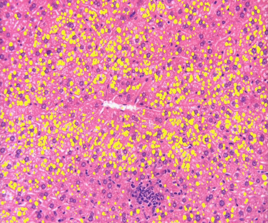
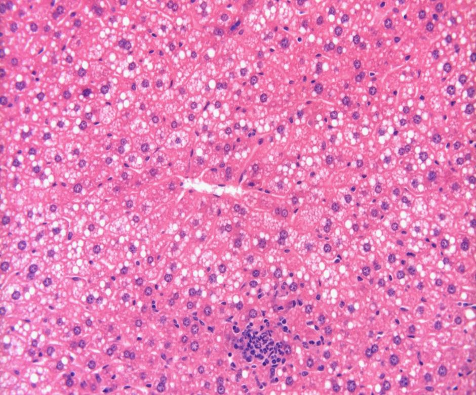
Hepatic steatosis quantification
- Quantification of histomorphological parameters in NAFLD rodent models .
- Accurate detection and quantification of fatty vacuoles in H&E stained rodent liver sections .
- Differentiation of fatty vacuoles from lumina of blood vessels and bile ducts .
- Strong and significant correlation (r = 0.87, p < .001) with semi-quantitative/manual assessment methods .
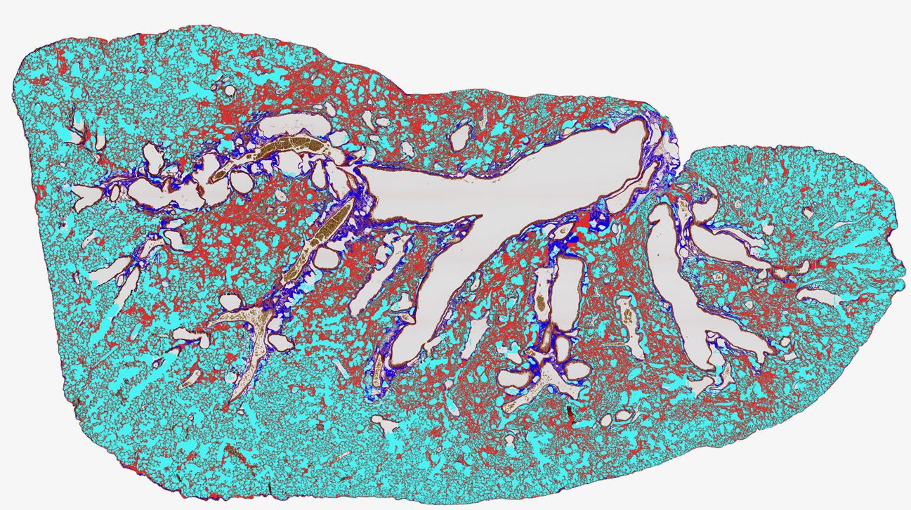
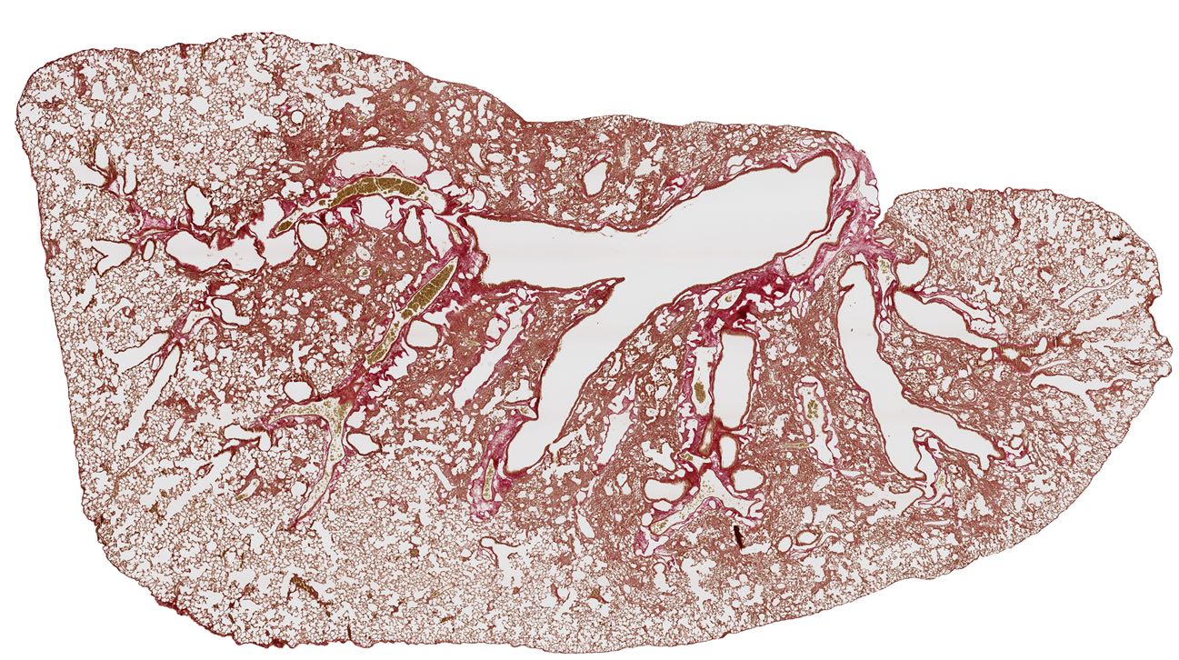
Pulmonary Fibrosis Grading
- Deep learning and Image processing network to quantify morphological changes in animal models for Bleomycin induced lung fibrosis .
- Accurate and objective segmentation and quantification of histomorphological parameters in rodent lung sections stained with Picrosirius Red and Haematoxyin & Eosin (H&E) .
- High throughput method for quantification of fibrosis in whole slide images of rodent lung.
- No restriction on field of analysis, negating selection bias to improve accuracy.
- Segmentation accuracy of more than 95% across parameters with strong correlation with manual grading using modified Ashcroft’s score.
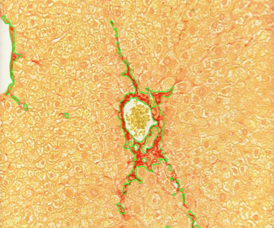
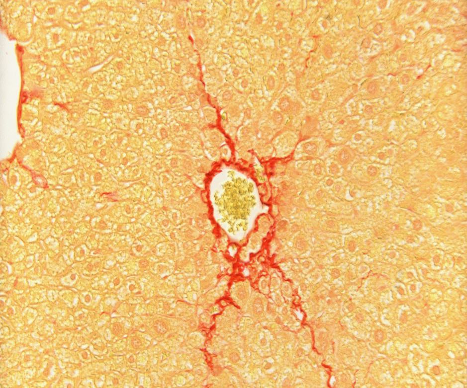
Hepatic Fibrosis Quantification
- Accurate quantification of liver fibrosis to aid evaluation of efficacy models for novel therapies.
- Segmentation and quantification of fibrous septa in Picrosirius stained sections of rodent liver.
- Applicable on microscope based analysis of field of view images as well as whole slide images.
- Strong positive correlation (p < .001) with semi-quantitative assessment by pathologists (Ishak score).
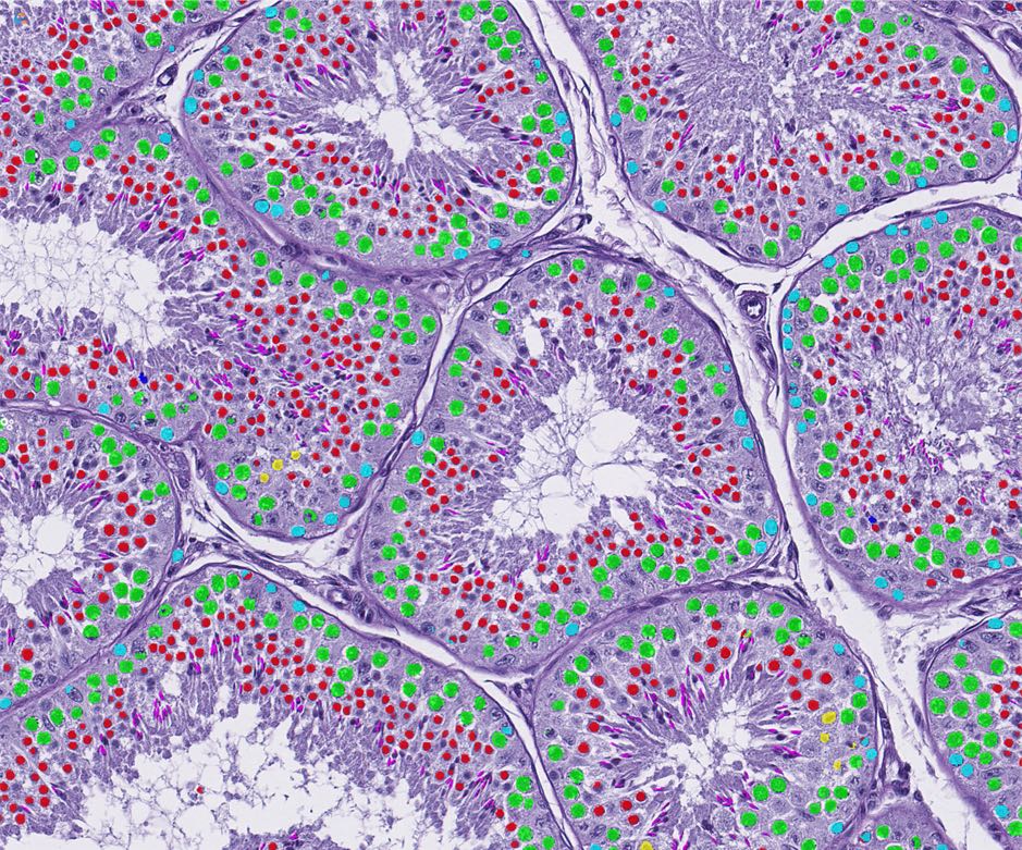
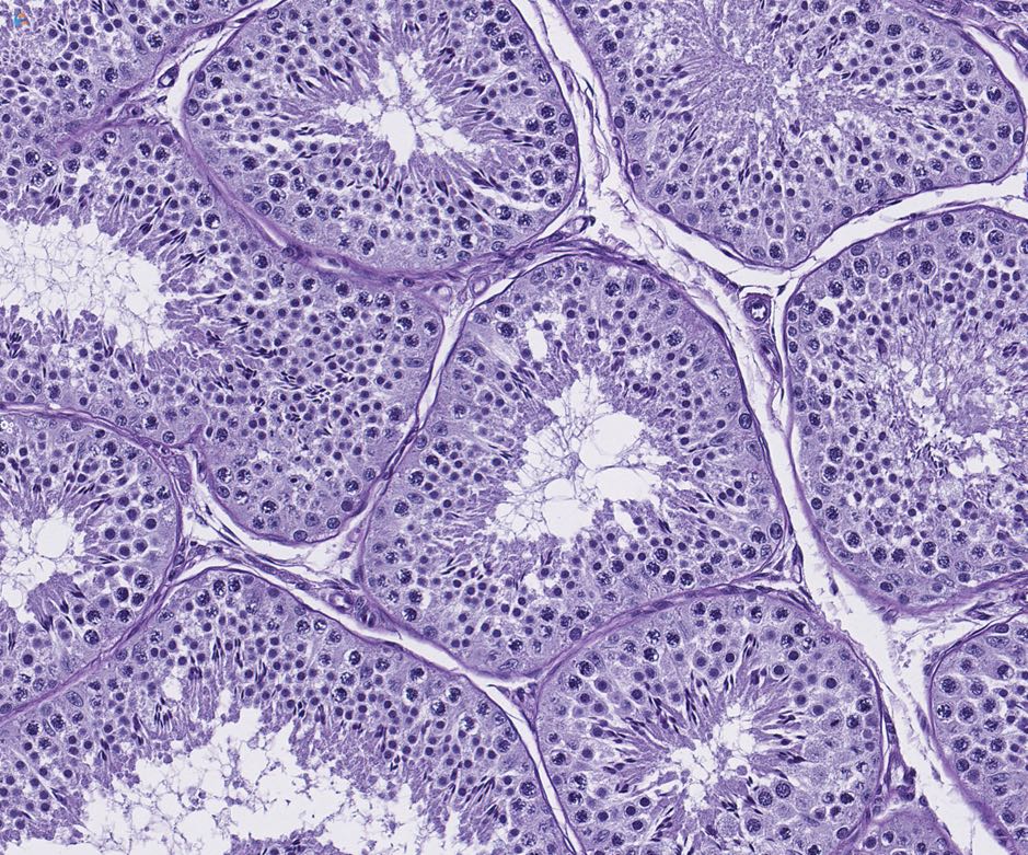
Spermatogenic staging assessment
- Automated method to identify the stages of rat spermatogenic cycle in digital images of Hematoxylin and Eosin stained testes.
- Negates the need for assessment of conventional PAS stained sections.
- Aids “stage-aware” histopathological evaluation of testes to detect effects on spermatogenesis in pre-clinical toxicology studies.
- Uses multiple criteria, based largely on the shape and movement of the elongating spermatids within the seminiferous tubule.
- Identifies spermatogenic stage of each tubule, allowing the pathologist to quickly evaluate the testis in a stage aware manner with rapid calculation of the stage frequencies.
- WSI staged in minutes as opposed to several days for manual staging; with objective, quantitative and reproducible outputs.
- Very good agreement for all the stages with Precision, Recall, and F1-score of 0.93 and 0.96 and 0.94 respectively, and overall accuracy of the staging analysis of 0.98.
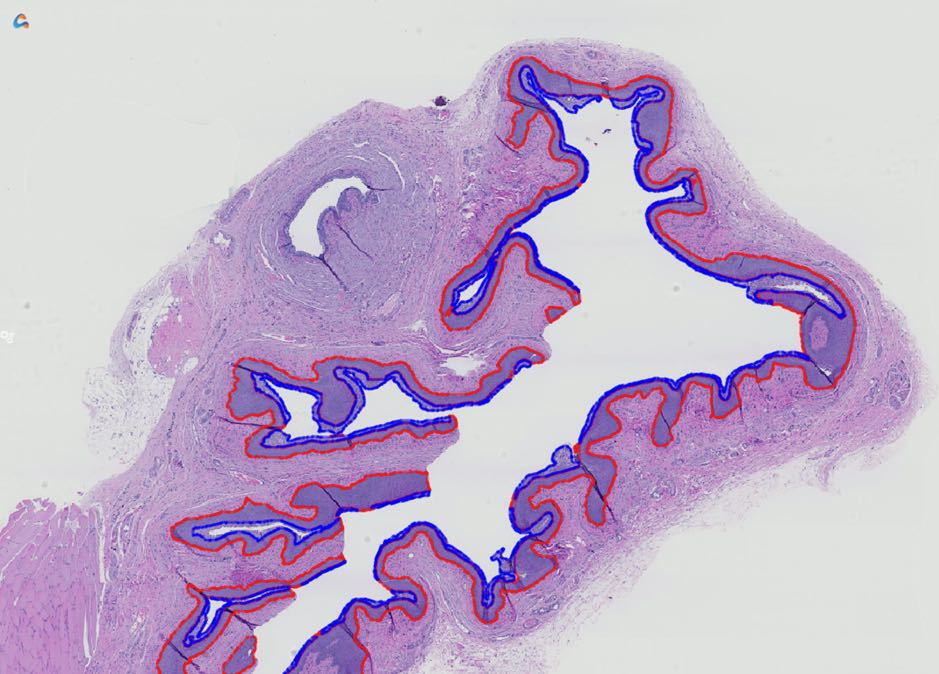
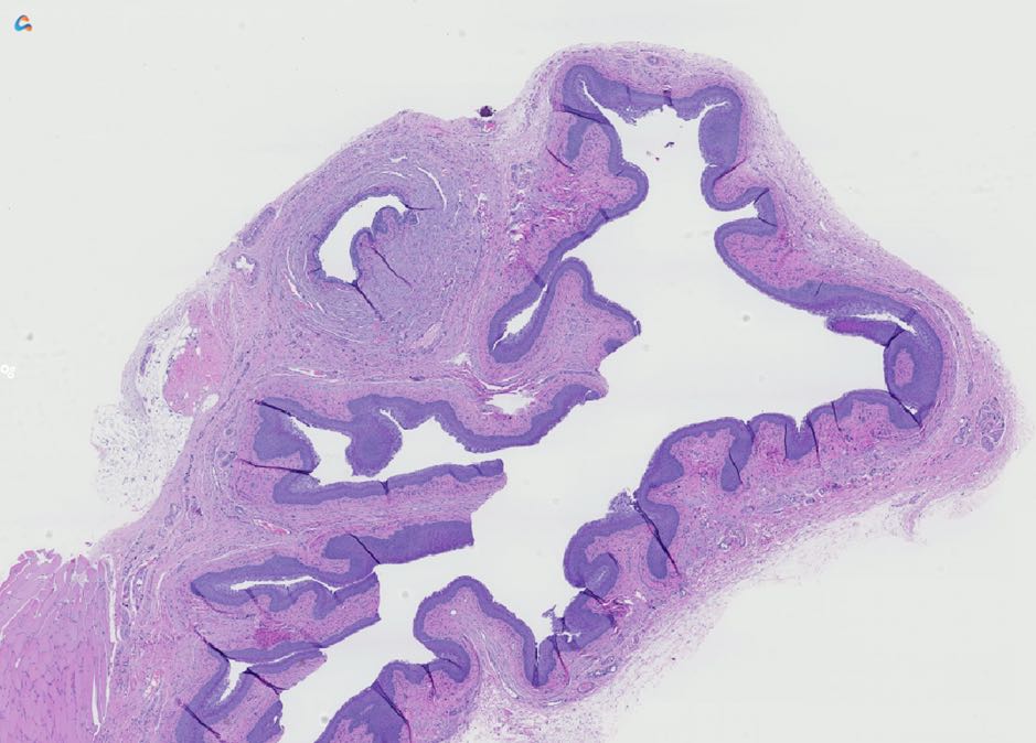
Estrous Staging Assessment
- Deep Learning and Image Processing solution for automated Estrous staging in rodents.
- Algorithms for evaluation of rodent vaginal and uterine sections, to aid stage aware assessment of female reproductive tissues.
- Segments and quantifies different histological layers, cellular changes and inflammatory cells.
- Sensitivity and Specificity range from 88% to 97% across stages (vaginal sections).
NOTE: Unless otherwise stated, all products are “Research Use Only”; not for use in diagnostic procedures.